


On the 236th birth anniversary of French physician René Laennec, considered the inventor of the stethoscope with a special doodle commemorating him on

René Laennec stumbled upon the design of the stethoscope while treating a female patient in 1816 at the Necker-Enfants Malades Hospital in Paris.
According to Laennec's own account, the young woman in question seemed to be suffering from what he described as symptoms of diseased heart. Unable to use the regular method of diagnosis in such a case (as the common method of putting the ear to the chest would be inapplicable in the case of the patient's age and gender), René Laennec recollected a simple and well-known fact in acoustics, the great distinctness with which we hear the scratch of a pin at one end of a piece of wood on applying our ear to the other. He rolled up a piece of paper into a cylinder and applied one end of it to the region of the heart and the other to his ear and was pleased to hear the action of the heart distinctly.
ADVERTISEMENT
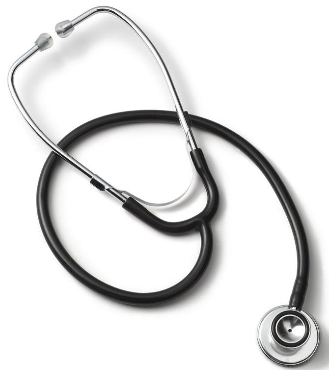
A stethoscope. Pic for representational purposes
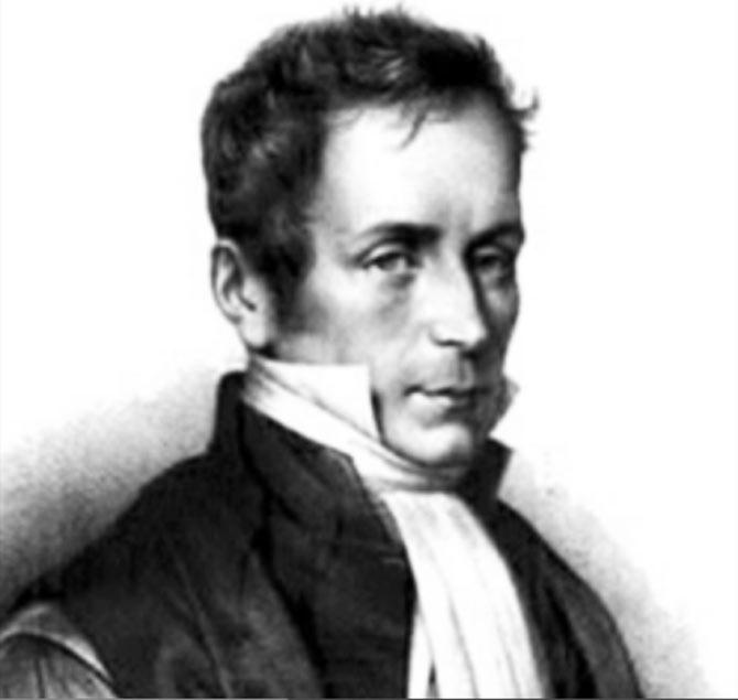
René Laennec. Pic/YouTube
Laennec built his first stethoscope as a 25 cm by 2.5 cm hollow wooden cylinder, which he later refined to comprise three detachable parts. His clinical work allowed him to follow chest patients from bedside to the autopsy table. He was therefore able to correlate sounds captured by his new instruments with specific pathological changes in the chest, in effect pioneering a new non-invasive diagnostic tool. Laennec was the first to classify and discuss the terms rales, rhonchi, crepitance, and egophony – terms that doctors now use on a daily basis during physical exams and diagnoses.
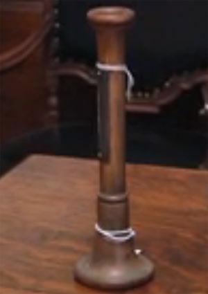
René Laennec's stethoscope. Pic/YouTube
After Laennec, Irish physician Arthur Leared invented the modern binaural stethoscope with two ear pieces in 1851 and George Cammann perfected the design of the instrument for commercial production in 1852, which has become the standard ever since.
The stethoscope, an acoustic medical device is used to listen to lung and heart sounds, intestines, blood flow in arteries and veins.
A look at other medical instruments commonly used today in medicine...
Sphygmomanometer: The sphygmomanometer, also known as a blood pressure meter or blood pressure gauge, was invented by Samuel Siegfried Karl Ritter von Basch in 1881. A more simplified and easily usable version by introduced by Scipione Riva-Rocci and the device was modernised and popularised within the medical community by Harvey Cushing in 1901.
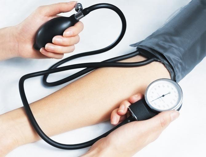
A sphygmomanometer. Picture for representational purposes
A sphygmomanometer is a medical instrument that measures blood pressure. It consists of an inflatable cuff, a measuring unit (the mercury manometer, or aneroid gauge), and a mechanism for inflation which may be a manually operated bulb and valve or a pump operated electrically. The usual unit of measurement of blood pressure is millimeters of mercury (mmHg) as measured directly by a manual sphygmomanometer. It is always used in conjunction with a means to determine at what pressure blood flow is just starting, and at what pressure it is unimpeded.
By observing the mercury in the column while releasing the air pressure with a control valve, one can read the values of the blood pressure in mm Hg. The peak pressure in the arteries during the cardiac cycle is the systolic pressure, and the lowest pressure (at the resting phase of the cardiac cycle) is the diastolic pressure. Measurement of the blood pressure is carried out in the diagnosis and treatment of hypertension (high blood pressure), and in many other healthcare scenarios.
Types
Manual sphygmomanometers: Used by trained practitioners, it is possible to obtain a basic reading through palpation alone, but this only yields the systolic pressure. Manual sphygmomanometers require a stethoscope for auscultation i.e. listening to the internal sounds of the body.
Mercury sphygmomanometers: These sphygmomanometers measure blood pressure by observing the height of a column of mercury; once made, errors of calibration cannot occur. They are often required in clinical trials of pharmaceuticals and for clinical evaluations of determining blood pressure for high-risk patients including pregnant women due to their high accuracy.
Aneroid sphygmomanometers: These sphygmomanometers are mechanical ones with a dial and require calibration checks, unlike mercury manometers. They are inexpensive and considered safer than the mercury based ones but are less accurate. A major cause of departure from calibration is mechanical jarring. Aneroids mounted on walls or stands are not susceptible to this particular problem.
Digital sphygmomanometers: These instruments use measurements and electronic calculations rather than auscultation and are easy to operate without training and can be used in noisy environments. Most instruments also display pulse rate. Digital instruments may use a cuff placed, in order of accuracy and inverse order of portability and convenience, around the upper arm, the wrist, or a finger. Some instruments claim also to measure arterial stiffness and irregular heartbeat. However such machines are not recommended for regular users as machines that claim to have 3 per cent accuracy rate, are usually inaccurate to over 7 per cent, and even provided two different readings when checked at the same time.
Tuning fork: This instrument was the invention of British musician John Shore. It is generally used by doctors to test for deafness and to categorize it. Tuning forks, usually C512, are used by medical practitioners to assess a patient's hearing. This is most commonly done with two exams called the Weber test and Rinne test, respectively. Lower-pitched ones (usually C128) are also used to check vibration sense as part of the examination of the peripheral nervous system.
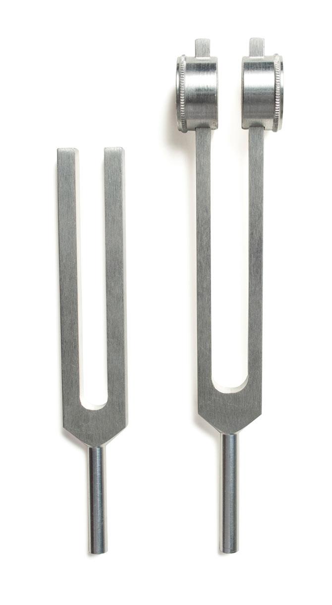
A couple of tuning forks. Picture for representational purposes
The Weber test, named after Ernst Heinrich Weber, is a quick screening test for hearing. It can detect unilateral (one-sided) conductive hearing loss (middle ear hearing loss) and unilateral sensorineural hearing loss (inner ear hearing loss).
The Rinne test, named after German otologist Heinrich Adolf Rinne, is primarily for evaluating loss of hearing in one ear and compares perception of sounds transmitted by air conduction to those transmitted by bone conduction through the mastoid. Thus, one can quickly screen for the presence of conductive hearing loss.
Ophthalmoscope: An ophthalmoscope is done as part of an eye examination, also known as an ophthalmoscopy or a fundoscopy and may be done as part of a routine physical examination. It is crucial in determining the health of the retina and the vitreous humor. Although, Charles Babbage is partially credited for its invention in 1847, Hermann von Helmholtz's 1851 reinvention enabled the usefulness of the ophthalmoscope to the medical profession.
An ophthalmoscope in use during an ophthalmoscopy. Picture for representational purposes
The instrument was later made into a hand-held device with concave mirror by Greek ophthalmologist Andreas Anagnostakis during his training in France. Francis A. Welch and William Noah Allyn invented the world's first hand-held direct illuminating ophthalmoscope in 1915.
Types
There are two types of ophthalmoscopes namely...
Direct ophthalmoscope: Commonly used during routine medical check-ups, its the size of a small flashlight with several lenses that can magnify upto 15 times.
Indirect ophthalmoscope: It can be either monocular or binocular and is used for peripheral viewing of the retina. Consisting of a light attached to a headband, a small handheld lens, it provides a wider view of the inside of the eye and thus enables a better view of the fundus even if the lens is clouded by cataracts.
Ophthalmoscopy enables the detection and evaluation of symptoms of various retinal vascular diseases or eye diseases such as glaucoma. Disorders like intracranial pressure, hydrocephalus, benign intracranial hypertension and even brain tumours can be discovered through this method if symptoms like headaches, swollen optic discs, or papilledema are detected during the ophthalmoscopy.
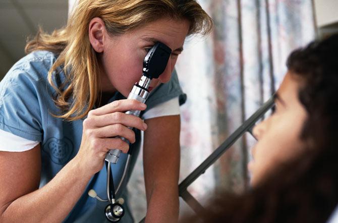
Otoscope: Also known as an auriscope, it is a medical device used to screen for illness during regular check-ups and also to investigate ear symptoms. An otoscope potentially gives a view of the ear canal and tympanic membrane, or eardrum. Since the eardrum is the border separating the external ear canal from the middle ear, its characteristics can be indicative of various diseases of the middle ear space. Diseases which may be diagnosed by an otoscope include otitis media and otitis externa, infection of the middle and outer parts of the ear, respectively.
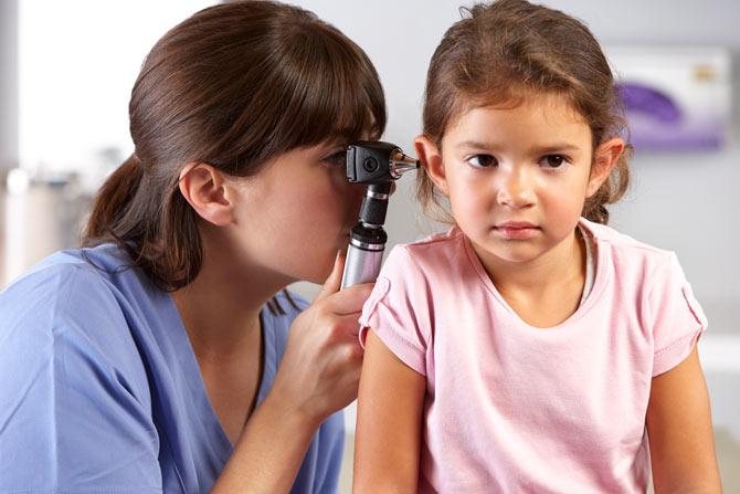
A doctor examines a patient's ear using an otoscope. Picture for representational purposes
Otoscopes are also frequently used for examining patients' noses (avoiding the need for a separate nasal speculum) and (with the speculum removed) upper throats. Many otoscopes used in doctors offices are wall-mounted, while others are portable. Wall-mounted otoscopes are attached by a flexible power cord to a base, which serves to hold the otoscope when it's not in use and also serves as a source of electric power, being plugged into an electric outlet. Portable models are powered by batteries in the handle; these batteries are usually rechargeable and can be recharged from a base unit.
The most commonly used otoscopes--those used in emergency rooms, pediatric offices, general practice, and by internists- are monocular devices. They provide only a two dimensional view of the ear canal, its contents, and usually at least a portion of the eardrum, depending on what is within the ear canal and its status.
 Subscribe today by clicking the link and stay updated with the latest news!" Click here!